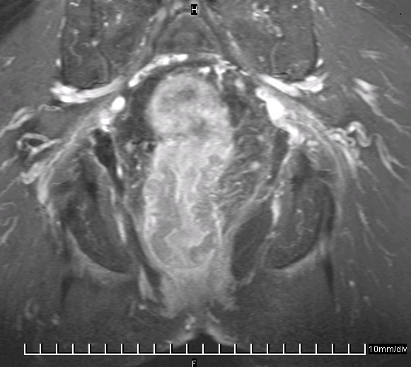
Gastro - Intestinale Tumore
Allgemeines

Karzinome

Diagnostik
| Tumor | niedriges T-Stadium | hohes T-Stadium | lokale Lymphknoten | distante Lymphknoten | M-Stadium |
| Ösophagus | EUS | PET-CT | PET/EUS | PET | PET |
| Magen | EUS | PET-CT | PET/EUS | PET | PET |
| Dünndarm | EUS/Kapsel | PET-CT | PET/EUS | PET | PET |
| Kolon | EUS | PET-CT | PET/EUS | PET | PET |
| Rektum | EUS | MRI | MRI | PET | PET |
| Leber | MRI/CT | MRI/CT | MRI/CT | MRI/CT | |
| Pankreas | MRI/CT | MRI/CT | MRI/CT | CT | |
| Biliarär | MRI | MRI | MRI/CT | CT |
Teil von
Quellen
Use of Imaging for GI Cancers.
J Clin Oncol 2015;33:1729-1736
2.) Sun W:
Multidisciplinary management of gastrointestinal cancers.
World Scientific 2016
3.) Messmann H, et al.:
Gastrointestinale Onkologie.
Thieme 2017
4.) Nagtegaal ID, Odze RD, Klimstra D et al.:
The 2019 WHO classification of tumours of the digestive system.
Histopathology 2020;76(2):182-8
5.) Torbenson MN, Ng IOL, Park YN, Roncalli M, Sakamato M
WHO classification of digestive systems tumors.
In: WHO Classification of Tumours,
2019, 5th ed, vol 1 International Agency for Research on Cancer, Lyon